[ad_1]
Summary: The activity of different populations of neurons in the amygdala governs whether mice interact with their peers, or indulge in self-centered behaviors.
Source: FMI
The brain’s emotion-processing center—the amygdala—is one of several brain regions involved in social behavior. But the exact role that this almond-shaped structure plays in the so-called ‘social brain’ remains mysterious.
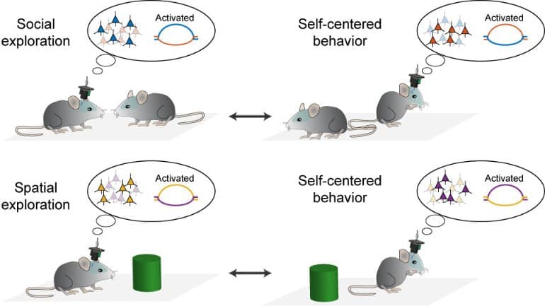
Now, researchers at the Friedrich Miescher Institute for Biomedical Research (FMI) in Basel, Switzerland, have found that the activity of different populations of neurons in the amygdala reflects whether mice interact with their peers, or whether they focus on self-centered behaviors such as grooming.
The findings, published in Nature, could help to understand how the activity pattern of groups of neurons sets an overall brain state, and how that influences behavior—including social interaction and other behaviors that are impaired in neuropsychiatric conditions such as autism and social anxiety.
State of mind
The brain fluctuates between distinct internal states, including hunger, anxiety and aggression, each of which can alter an animal’s behavior. For example, a hungry mouse will move around and look for food, whereas an anxious one will freeze in place or only hesitantly explore the surrounding environment, says study co-senior author and FMI senior group leader Andreas Lüthi. However, Lüthi adds, the networks of neurons that encode different brain states remain unclear.
In a 2019 study, Lüthi’s team found that two large sets of neurons in the mouse amygdala showed opposing patterns of activity when the animals switched between exploring the environment or performing defensive behaviors such as freezing—a finding that hinted at a key role of the amygdala in coordinating brain states.
But the researchers felt there was more to the story. Studies in monkeys have suggested that the amygdala is important for social behavior, and altered amygdala structure or function has been linked to a series of neuropsychiatric conditions that affect social interaction, from autism spectrum disorders to schizophrenia. So, Maria Sol Fustiñana and Yael Bitterman from Lüthi’s group and their colleagues at the FMI and the Novartis Institutes for BioMedical Research (NIBR) set out to look at neuronal activity in the amygdala when two mice were free to interact in a Plexiglas chamber.
“As experimenters, we took a backseat and let the animals decide when they wanted to engage with another mouse placed in the chamber, for how long and in what manner,” says Bitterman, who is co-senior author on the study.
Ready to mingle
The researchers recorded videos of the mice’s social encounters and developed an algorithm that characterized the animals’ behavior based on their video tracking. Mice rarely showed aggressive behaviors; instead, they sought to make each other’s acquaintance or, in some cases, avoided interacting with the stranger.
To track the activity of nearly one hundred neurons in the amygdala, the researchers used tiny head-mounted microscopes that peered deep into the brains of mice without interfering with the animals’ normal behavior.
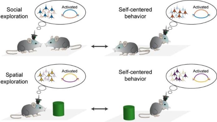
The team found that one population of neurons was active only when the mice engaged in social behavior—no matter if they peacefully interacted or they tried to avoid the other animal. A second group of brain cells in the amygdala became active when the mice switched to self-grooming or other non-social behaviors.
Similarly, the team found two populations of amygdala neurons that displayed opposing activation when a single mouse put in a Plexiglas chamber switched between grooming and exploring the chamber or an inanimate object. But the neurons becoming activated as the solitary mouse explored the environment were not the same as those active when the animal interacted with another mouse.
When two mice were in the same chamber, their attention was mostly focused on the stranger: although the rodents typically explored their surroundings while not engaging in social interaction, they didn’t wander around the whole chamber. Under these conditions, the researchers observed that amygdala neurons preferentially code for social interaction rather than spatial exploration.

“The top priority is the other animal, probably because it could be a threat or a potential mate,” Lüthi says. He notes that many people think of the amygdala as the brain’s anxiety detector. Instead, he says, “the social engagement signal covers both avoidance and approach.”
Figuring out how the amygdala processes such different behaviors was “absolutely rewarding,” says Fustiñana, who is lead author on the study and was part of a joint FMI-Novartis postdoctoral program that fostered interactions between the FMI and NIBR. “This entire project was a great challenge—from establishing a naturalistic behavioral experiment with two freely moving mice to imaging a deep brain structure and doing the required computational analyses,” she says. “This wouldn’t have been possible without a concerted team effort.”
Next, the researchers plan to look at how internal state signals are broadcast across the brain, and whether manipulating amygdala neurons encoding brain states could lead to changes in behavior.
Because psychiatric disorders are accompanied by altered brain states, Lüthi says, understanding how the brain switches between states might help to figure out what goes awry in those disorders and identify potential strategies to alleviate dysfunctional behaviors.
About this social neuroscience research news
Source: FMI
Contact: Press Office – FMI
Image: The image is credited to FMI
Original Research: Closed access.
“State-dependent encoding of exploratory behaviour in the amygdala” by Maria Sol Fustiñana et al. Nature
Abstract
State-dependent encoding of exploratory behaviour in the amygdala
The behaviour of an animal is determined by metabolic, emotional and social factors. Depending on its state, an animal will focus on avoiding threats, foraging for food or on social interactions, and will display the appropriate behavioural repertoire. Moreover, survival and reproduction depend on the ability of an animal to adapt to changes in the environment by prioritizing the appropriate state. Although these states are thought to be associated with particular functional configurations of large-brain systems, the underlying principles are poorly understood.
Here we use deep-brain calcium imaging of mice engaged in spatial or social exploration to investigate how these processes are represented at the neuronal population level in the basolateral amygdala, which is a region of the brain that integrates emotional, social and metabolic information.
We demonstrate that the basolateral amygdala encodes engagement in exploratory behaviour by means of two large, functionally anticorrelated ensembles that exhibit slow dynamics. We found that spatial and social exploration were encoded by orthogonal pairs of ensembles with stable and hierarchical allocation of neurons according to the saliency of the stimulus.
These findings reveal that the basolateral amygdala acts as a low-dimensional, but context-dependent, hierarchical classifier that encodes state-dependent behavioural repertoires. This computational function may have a fundamental role in the regulation of internal states in health and disease.
[ad_2]
Source link


