

[ad_1]
Summary: Neurons from brain tissue samples of people with schizophrenia showed greater deviation, both from neurons from samples of people without the condition and from others with schizophrenia. Researchers say the structure of the neurons is unique to each individual with schizophrenia.
Source: DOE/Argonne National Laboratory
Schizophrenia, a chronic, neurological brain disorder, affects millions of people around the world. It causes a fracture between a person’s thoughts, feelings and behavior. Symptoms include delusions, hallucinations, difficulty processing thoughts and an overall lack of motivation. Schizophrenia patients have a higher suicide rate and more health problems than the general population, and a lower life expectancy.
There is no cure for schizophrenia, but the key to treating it more effectively is to better understand how it arises. And that, according to Ryuta Mizutani, professor of applied biochemistry at Tokai University in Japan, means studying the structure of brain tissue. Specifically, it means comparing the brain tissues of schizophrenia patients with those of people in good mental health, to see the differences as clearly as possible.
“The current treatment for schizophrenia is based on many hypotheses we don’t know how to confirm,” Mizutani said. “The first step is to analyze the brain and see how it is constituted differently.”
To do that, Mizutani and his colleagues from several international institutions collected eight small samples of brain tissue — four from healthy brains and four from those of schizophrenia patients, all collected post-mortem — and brought them to beamline 32-ID of the Advanced Photon Source (APS), a U.S. Department of Energy (DOE) Office of Science User Facility at DOE’s Argonne National Laboratory.
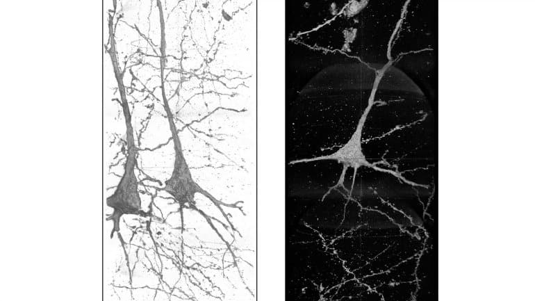
At the APS, the team used powerful X-rays and high-resolution optics to capture three-dimensional images of those tissues. (Researchers collected similar images at the Super Photon Ring 8-GeV [SPring-8] light source facility in Japan.) The resolution of the X-ray optics used at the APS can be as high as 10 nanometers. That’s about 700 times smaller than the width of the average red blood cell, and there are five million of those cells in a drop of blood.
“There are only a few places in the world where you can do this research,” Mizutani said. “Without 3D analysis of brain tissues this work would not be possible.”
According to Vincent De Andrade, physicist in Argonne’s X-ray Science Division, capturing images at high resolution presents a challenge, since the neurons being imaged can be centimeters long. The neuron is the basic working unit of the brain, a cell within the nervous system that transmits information to other cells to control body functions. The human brain has roughly 100 billion of these neurons, in various sizes and shapes.
“The sample has to move through the X-ray beam to trace the neurons through the sample,” De Andrade explained. “The field of view of our X-ray microscope is about 50 microns, about the width of a human hair, and you need to follow these neurons over several millimeters.”
What these images showed is that the structures of these neurons are uniquely different in each schizophrenia patient, which Mizutani said is evidence that the disease is associated with those structures. Images of healthy neurons were relatively similar, while neurons from schizophrenia patients showed far more deviation, both from the healthy brains and from each other.
More study is needed, Mizutani said, to figure out exactly how the structures of neurons are related to the onset of the disease and to devise a treatment that can alleviate the effects of schizophrenia. As X-ray technology continues to improve — the APS, for example, is scheduled to undergo a massive upgrade that will increase its brightness up to 500 times — so will the possibilities for neuroscientists.
“The APS upgrade will allow for better sensitivity and resolution for imaging, making the process of mapping neurons in the brain faster and more precise,” De Andrade said. “We would need resolutions of better than 10 nanometers to capture synaptic connections, which is the holy grail for a comprehensive mapping of neurons, and those should be achievable with the upgrade.”
De Andrade also noted that while electron microscopy has been used to map the brains of small animals — fruit flies, for instance — that technique would take a long time to image the brain of a larger animal, such as a mouse, let alone a full human brain. Ultrabright, high energy X-rays like those at the APS, he said, could speed up the process, and advances in technology will help scientists get a more complete picture of brain tissue.
For neuroscientists like Mizutani, the end goal is fewer people suffering with brain diseases like schizophrenia.
“The differences in brain structure between healthy and schizophrenic people must be linked to mental disorders,” he said. “We must find some way to make people healthy.”
About this schizophrenia research news
Source: DOE/Argonne National Laboratory
Contact: Beth Schlesinger – DOE/Argonne National Laboratory
Image: The image is credited to Ryuta Mizutani
Original Research: Open access.
“Structural diverseness of neurons between brain areas and between cases” by Ryuta Mizutani, Rino Saiga, Yoshiro Yamamoto, Masayuki Uesugi, Akihisa Takeuchi, Kentaro Uesugi, Yasuko Terada, Yoshio Suzuki, Vincent De Andrade, Francesco De Carlo, Susumu Takekoshi, Chie Inomoto, Naoya Nakamura, Youta Torii, Itaru Kushima, Shuji Iritani, Norio Ozaki, Kenichi Oshima, Masanari Itokawa& Makoto Arai. Translational Psychiatry
Abstract
See also

Structural diverseness of neurons between brain areas and between cases
The cerebral cortex is composed of multiple cortical areas that exert a wide variety of brain functions. Although human brain neurons are genetically and areally mosaic, the three-dimensional structural differences between neurons in different brain areas or between the neurons of different individuals have not been delineated.
Here we report a nanometer-scale geometric analysis of brain tissues of the superior temporal gyrus of schizophrenia and control cases. The results of the analysis and a comparison with results for the anterior cingulate cortex indicated that (1) neuron structures are significantly dissimilar between brain areas and that (2) the dissimilarity varies from case to case. The structural diverseness was mainly observed in terms of the neurite curvature that inversely correlates with the diameters of the neurites and spines.
The analysis also revealed the geometric differences between the neurons of the schizophrenia and control cases. The schizophrenia cases showed a thin and tortuous neuronal network compared with the controls, suggesting that the neuron structure is associated with the disorder. The area dependency of the neuron structure and its diverseness between individuals should represent the individuality of brain functions.
[ad_2]
Source link


 – tadalafil – Erectile Dysfunction-Beemedz.com")


